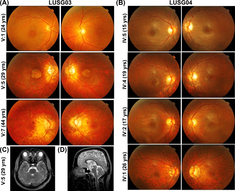
Figure 2.
Color fundus photography of the right and left macula, respectively, of individuals with cone dystrophy from families LUSG03 (A) and LUSG04 (B). Myopic fundus changes were variably present in LUSG03 V:5, V:7 (A) and LUSG04 IV:1, IV:4, and IV:5 (B), including tilted optic disc, alpha zone atrophy, mild temporal disc pallor, and parapapillary RPE thinning consistent with early staphyloma formation. Macular atrophy was observed in LUSG03 V:5 and bull's eye lesions are visible in LUSG03 V:1, V:7 and LUSG04 IV:1, IV:2, and IV:4. Brain magnetic resonance imaging axial T2 image (C) and sagittal T2 image (D) of LUSG03 individual IV:5, demonstrating no increased interpeduncular fossa, normal cerebellar peduncles, and normal vermis.

