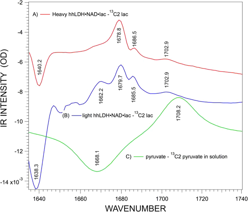
Figure 3.
Difference IR spectra of [12C]pyruvate minus [13C]pyruvate in (A) heavy hhLDH·NADH·pyruvate, (B) light hhLDH·NADH· pyruvate, and (C) pyruvate in solution. The starting concentrations of the reactants, enzyme, NAD, and lactate in parts A and B, were 7:7:80 mM. The prominent peak in the difference spectrum (panel B) of light protein at 1662 cm−1 is a subtraction artifact, since (1) its relative intensity varies from run to run while the other bands do not and (2) the band is not present in the heavy protein spectrum. See also the Supporting Information. The protein background spectrum of nonlabeled (light) protein rapidly increases at this point but is downshifted for the labeled (heavy) protein.

