Figure 1:

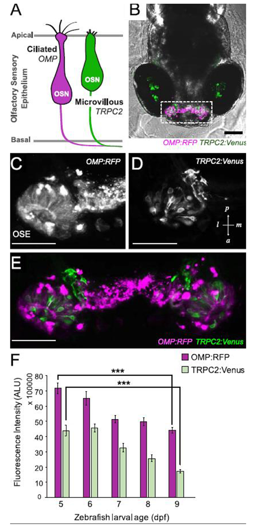
Two populations of olfactory sensory neurons (OSNs) are differentially labeled in double-transgenic zebrafish. A. Schematic of the two predominant OSNs in zebrafish olfactory sensory epithelium. B-E. In vivo confocal images of a double transgenic zebrafish larva (6 dpf, B) expressing OMP:RFP (C) in ciliated OSNs and TRPC2:Venus (D) in microvillous OSNs within the olfactory sensory epithelium (OSE; E). Images are stacked projections of optical sections. Scale bars = 100 μM (B); 50 μM (C-E). F. Semi-quantitative measurement of fluorescence intensity of OMP:RFP and TRPC2:Venus in OSE during early larval development between 5-9 dpf (measured in arbitrary light units, ALU) showing decrease in OSNs over time. Error bars = ± SE (standard error); n = 4-5 fish per time point. Asterisks indicate statistical significant change from 5 dpf larvae at ***p<0.001 significance levels.
