Figure 2:

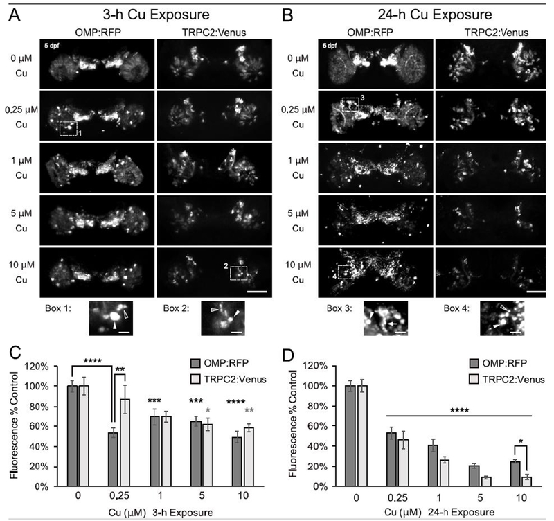
Severity of OSN damage by Cu is dose-dependent and time-dependent. A-B. Representative confocal images (stacked projections) of larval zebrafish OSE show changes in ciliated (OMP:RFP) and microvillous (TRPC2:Venus) OSNs following either a 3-h (A) or 24-h (B) exposure to Cu at 5 dpf. Magnification of numbered dotted boxes are shown below and highlight some of the observed morphological OSN changes such as dense cell bodies (white arrowheads), cell fragmentation (open arrowheads), and axonal retraction (white arrow). Scale bars = 50 μM and 10 μM (boxes). C-D. Semi-quantitative analysis of OMP:RFP and TRPC2:Venus fluorescence after (C) 3-h and (D) 24-h Cu exposure. Fluorescence intensity is normalized to age-matched non-exposed (0 μM Cu) controls. Error bars = ± SE; n=4-5 fish per condition. Asterisks indicate statistical significant change from 0 μM Cu control larvae, unless otherwise indicated (black asterisks= OMP:RFP; gray asterisks = TRPC2:Venus), at *p<0.05, **p<0.01, ***p<0.001, and ****p<0.0001 significance levels.).
