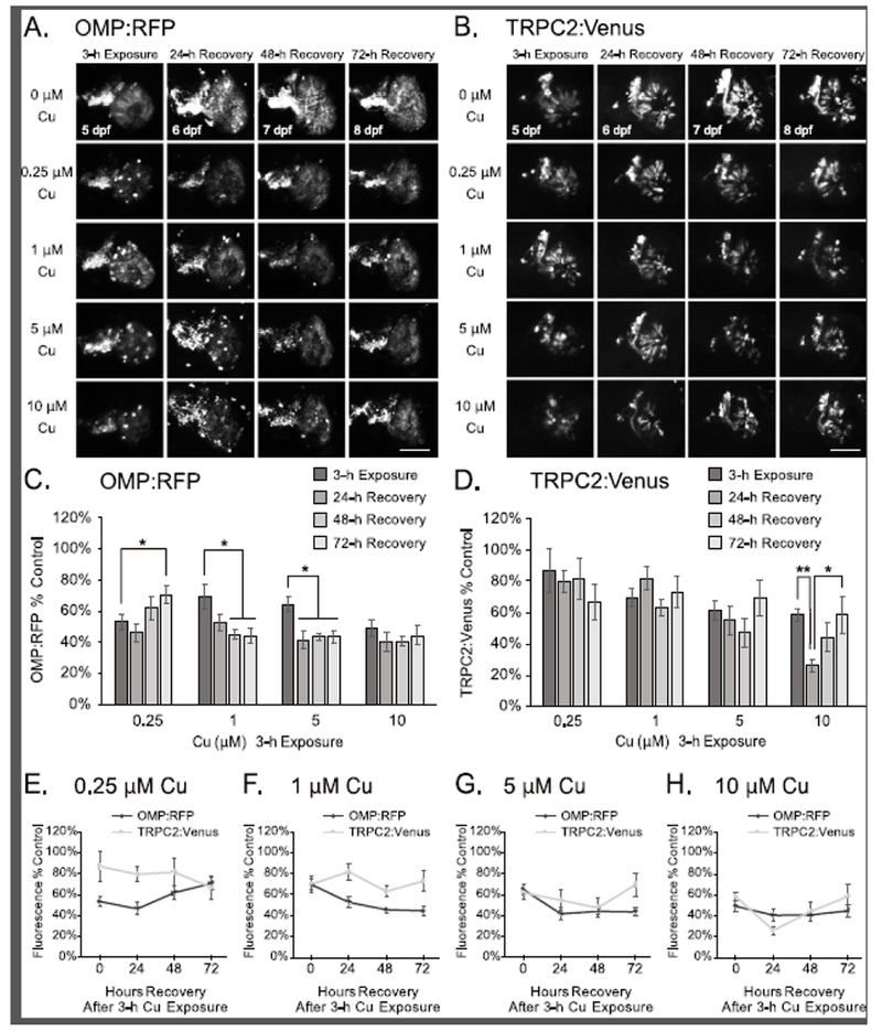
Figure 3:

Partial OSN regeneration is observed following recovery after 3-h Cu-induced injury at 5 dpf. A-B. Representative confocal images of a single OSE from double-transgenic larval zebrafish show OMP:RFP-labeled ciliated OSNs (A) and TRPC2:Venus-labeled microvillous OSNs (B) imaging after 3h exposure of Cu at different doses after 24, 48, and 72 hours of recovery. Scale bars = 50 μM. C-D. Semi-quantitative analysis of OMP:RFP (C) and TRPC2:Venus (D) fluorescence in confocal images. E-H. Graphs comparing OMP:RFP and TRPC2:Venus fluorescence at 0.25 μM (E), 1 μM (F), 5 μM (G), and 10 μM (H) Cu exposure for 3 h (same dataset as presented in Fig. 3C-D). Fluorescence intensity is normalized to the age-matched non-exposed (0 μM Cu) controls. Error bars = ± SE; n=4-5 fish per condition. Asterisks indicate statistically significant change at *p<0.05 and **p<0.01 significance levels.
