Figure 2:
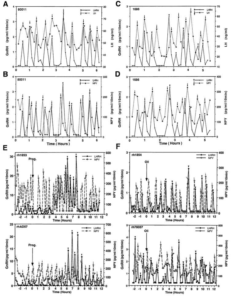
Correlated release of GnRH in the stalk median eminence and LH release in general circulation (A, C) and synchronous release of GnRH and NPY in the stalk median eminence (B, D) from an orchidectomized male monkey (left) and an ovariectomized female monkey (right). Arrowheads indicate LHRH pulses and arrows indicate LH or NPY pulses detected by PULSAR. Note that in male monkey, all GnRH peaks are synchronized with LH pulses (A), and 9 of 10 NPY peaks either precede by one point or coincide with corresponding GnRH peaks (C). Similarly, in female monkey, GnRH and LH pulses and NPY and GnRH pulses are highly correlated as GnRH peaks either precede or coincide with LH peaks (B), and NPY and GnRH pulses are also highly correlated (D). Effects of progesterone (E) or oil (F, control for progesterone) on the release of GnRH (solid line) and NPY (broken line) measured in aliquots of the same perfusate samples from the S-ME of two ovariectomized female monkeys. Progesterone or oil was injected at time zero in animals treated with EB (30 μg) 24 h earlier. Arrowheads and arrows indicate pulses of GnRH and NPY, respectively, detected by Pulsar. Note that in E the pulse frequency was increased by progesterone in both GnRH and NPY release, maintaining the synchronous release of both neuropeptides. In contrast, the pulse amplitude of GnRH release increased dramatically 3–9 h after progesterone treatment, while the pulse amplitude of NPY remained unaltered. In F, oil injection altered neither the pulse frequency nor pulse amplitude. Again, the synchronous release of GnRH and NPY was unaltered. Reproduced from Woller et al., Endocrinology. 130, 2333–2342, 1992, and Woller and Terasawa, Endocrinology 135, 1679–1686, 1994, with permission pending.

