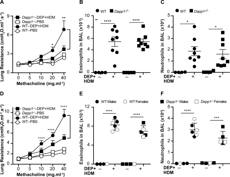
Fig 5. Functional validation of Dapp1 as a DEP-responsive gene.
A and D) Lung resistance after inhalation exposure to DEP and HDM is not increased in Dapp1-/- mice compared to WT animals. DEP/HDM exposure induced eosinophilia (B and E) and neutrophilia (C and F) to the same extent in BAL fluid from Dapp1-/- and WT mice. Mice were first sensitized on day 0 through a 100μl intraperitoneal injection containing 200μg DEP and 25μg HDM + 2.25mg Alum, as an adjuvant. On days 7–10, mice were placed in insulated chambers daily for 20mins supplied with ambient air and saturated with either aerosolized PBS or both 200μg DEP and 25μg HDM. On day 11, airway hyperreactivity was measured by invasive plethysmography, followed by collection of BAL. Cell counts in BAL fluid were determined by flow cytometry. WT control animals were either female C57BL/6J mice purchased from the Jackson Laboratories (A-C) or WT littermates of both sexes generated through an intercross between Dapp1+/- heterozygote mice (D-F). For both experiments, n = 4–8 mice in each group. Data are shown as mean ± SE. ****p<0.0001; ***p<0.001; **p<0.005; *p<0.05.

