Figure 4.
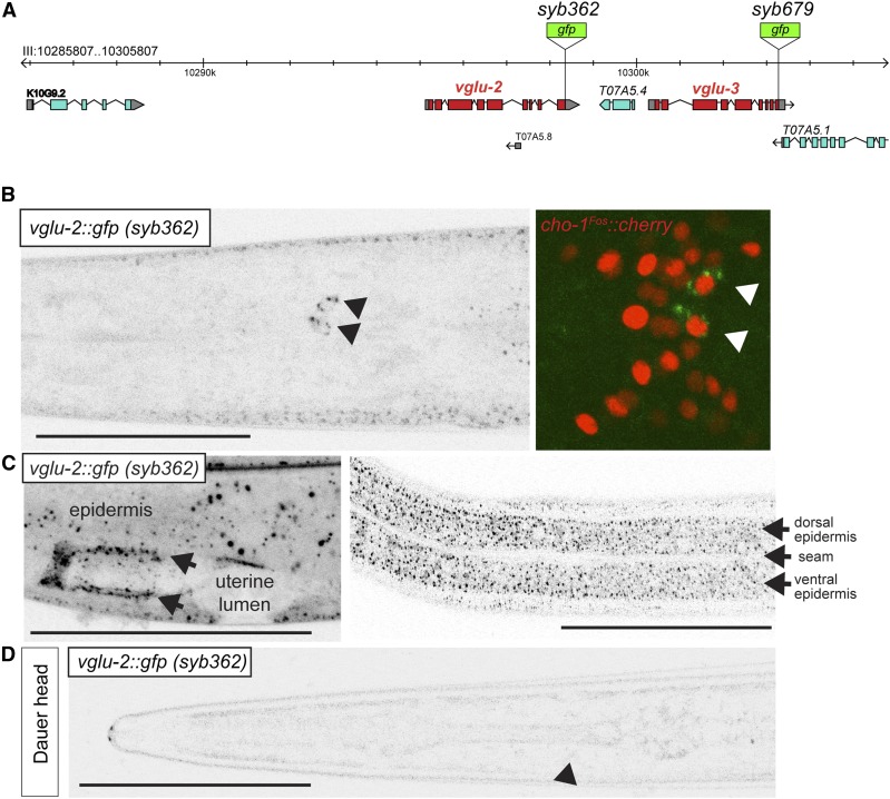
Expression and localization patterns of vglu-2 in the nervous system. (A) Schematic of vglu-2 reporter vglu-3 loci and location of the gfp cassettes generated by CRISPR/Cas9 genome engineering. (B–D) GFP expression in the vglu-2(syb362[vglu-2::gfp]) strain. (B) Ventral view of a young adult animal, showing neuronal, punctate expression in AIA neurons (marked with black arrowhead). AIA identity was confirmed by crossing reporter with AIA-expressed, nuclear-localized cho-1 reporter (right panel; red signal). Green signal in right panel is VGLU-2::GFP (marked with arrows). (C) Nonneuronal expression in epidermis and uterine tissue lining the uterine lumen (arrows). (D) Lateral view of a dauer-stage animal. Black arrowhead indicates approximate position of AIA, in which VGLU-2::GFP is downregulated, as it is in the epidermis. Bar, 50 μm.

