. 2020 Jan 3;9:e50065. doi: 10.7554/eLife.50065

Figure 2. UNC-3 has a dual role in cholinergic ventral cord motor neurons.

(A) Terminal identity markers of VD neurons (ser-2, oig-1) are ectopically expressed in unc-3-depleted MNs. Representative images of larval stage 4 (L4) hermaphrodites are shown. Similar results were obtained in adult animals. Arrowheads point to MN cell bodies with gfp marker expression. Green fluorescence signal is shown in white for better contrast. Dotted black box indicates imaged area. (B) Quantification of VD markers (ser-2, oig-1) in WT and unc-3 (n3435) at L4. N > 15. ***p<0.001. For details on box plots, see Materials and methods. (C) Terminal identity markers of VC neurons (ida-1, srb-16, glr-5) are ectopically expressed in unc-3-depleted MNs. Representative images of larval stage 4 (L4) hermaphrodites are shown. Similar results were obtained in adult animals. Arrowheads point to MN cell bodies with gfp marker expression. Green fluorescence signal is shown in white for better contrast. Dotted black box indicates imaged area. (D): Quantification of VC markers (ida-1, srb-16, glr-5) in WT and unc-3 (n3435) at L4. Individual data points are dot-plotted. N > 15. ***p<0.001. (E) Distinct MNs acquire VC-like or VD-like terminal identity features in unc-3 (n3435) mutants. The VC marker in green (ida-1::gfp) and the VD marker in red (ser-2::rfp) do not co-localize in WT or unc-3 (n3435) mutants. Representative images are shown. Individual VC/VC-like and VD/VD-like neurons are pointed and circled, respectively,(VD: dotted circles; VC: arrowheads) to highlight that an individual MN never expresses both markers. (F) Quantification of data shown in E. N > 16. (G) Schematic that summarizes the dual role of unc-3. Apart from activating cholinergic MN terminal identity genes, UNC-3 prevents expression of VD and VC terminal features in distinct cells (‘VD-like’ versus ‘VC-like’).

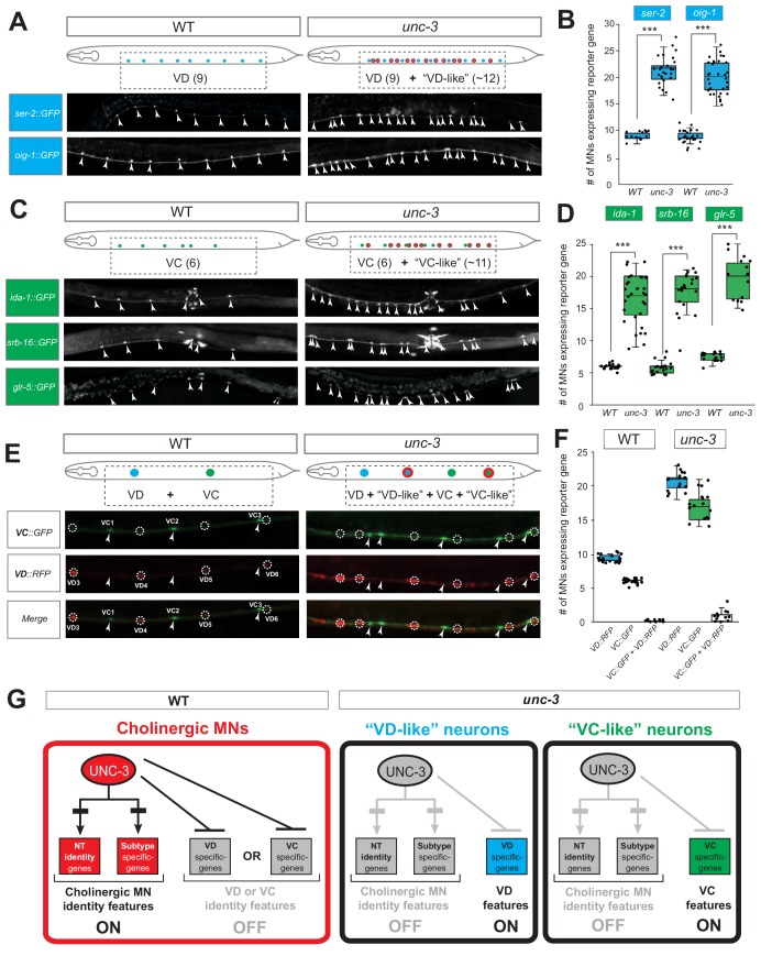
Figure 2.

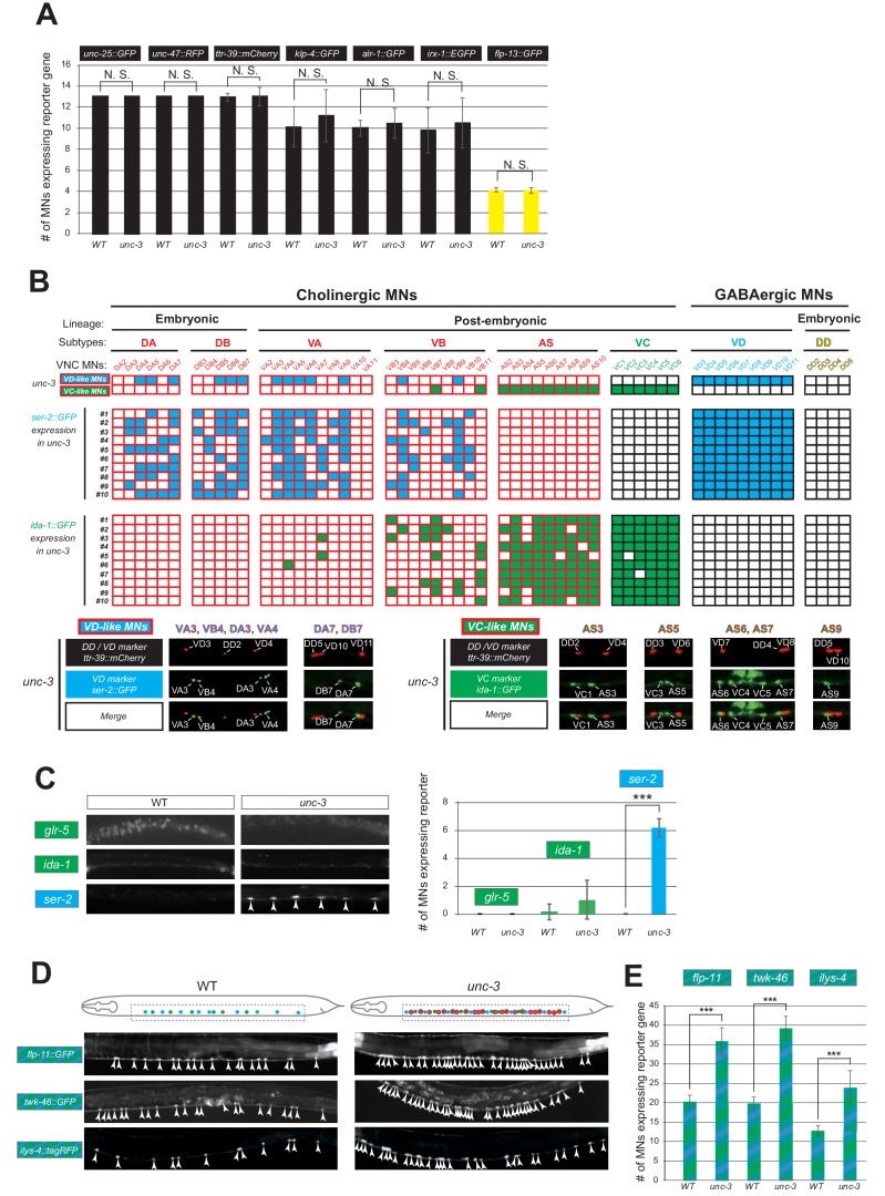
Figure 2—figure supplement 1. UNC-3 selectively prevents expression of VD and VC terminal identity features in distinct cholinergic MNs.

Figure 2—figure supplement 1.

(A) Quantification of six VD/DD (unc-25::gfp, unc-47::rfp, ttr-39::mCherry, klp-4::gfp, alr-1::gfp and irx-1::egfp) and one DD (flp-13::gfp) markers in WT and unc-3 (n3435) animals at L4. No statistically significant effects were observed in unc-3 mutants. N > 13. N. S: not significant. (B) A table showing that VD and VC identity genes are ectopically expressed in distinct cholinergic MNs upon unc-3 depletion. Top two rows: A summary map of VD and VC gene expression with single-cell resolution in unc-3 mutants, which is based on the co-localization analysis shown below. Each column represents an individual MN in the VNC of an unc-3-depleted hermaphrodite worm. Ten randomly selected worms that co-express VD/DD marker ttr-39::mCherry and VD marker ser-2::gfp or VC marker ida-1::gfp are analyzed. The expression of ttr-39::mCherry is not affected by unc-3 depletion, which serves as a positional landmark for reference. This landmark together with the invariant MN cell body position along the VNC enable the identification of distinct unc-3-depleted MNs that lose cholinergic features and concomitantly gain either ‘VD-like’ or ‘VC-like’ features. Below this table, examples of the co-localization analysis are provided. Representative magnified images are shown. The identity of unc-3-depleted MNs that acquire VC or VD terminal features is shown. Note that VC-like terminal features are mainly acquired by MNs of the AS cholinergic subtype, whereas VD-like terminal features are acquired by select members of four cholinergic MN subtypes (DA, DB, VA, VB). (C) The DA and DB cholinergic MNs are born embryonically and therefore present in the VNC at L1. Corroborating our results from panel B, only the VD terminal identity marker (ser-2) shows ectopic expression in DA and DB neurons of unc-3 mutants at L1. This is not the case for the VC terminal identity markers (glr-5, ida-1). Representative images are shown on the left. Arrowheads point to MN cell bodies with ectopic gfp marker expression. Green fluorescence signal is shown in white for better contrast. Quantification is on the right. N > 22. ***p<0.001. (D) Terminal identity markers of VD/VC MNs (flp-11, twk-46 and ilys-4) are ectopically expressed in unc-3-depleted cholinergic MNs. Representative images of larval stage 4 (L4) hermaphrodites are shown. Similar results were obtained in adult animals. Arrowheads point to MN cell bodies with marker expression. Fluorescence signal is shown in white for better contrast. Dotted black box indicates imaged area. (E) Quantification of data shown in panel D.. N > 18. ***p<0.001.
Figure 2—figure supplement 2. The dual role of UNC-3 in cholinergic MNs extends to both C. elegans sexes.

Figure 2—figure supplement 2.

(A) Quantification of four sex-shared cholinergic MN markers (unc-17, cho-1, acr-2, unc-129) expression in WT and unc-3 (n3435) male animals at L4. N > 13. ***p<0.001. (B) Quantification of VD marker (ser-2 and oig-1) expression in L4 stage WT and unc-3 (n3435) male animals. No significant difference was found between the number of MNs ectopically expressing each marker. N > 11. ***p<0.001. N. S: not significant. (C) Quantification of the male-specific CA markers (ida-1, srb-16 and glr-5) in young adult stage WT or unc-3 (n3435) male animals. N > 14. ***p<0.001. (D) Schematic summarizing the dual role of UNC-3 in C. elegans males.