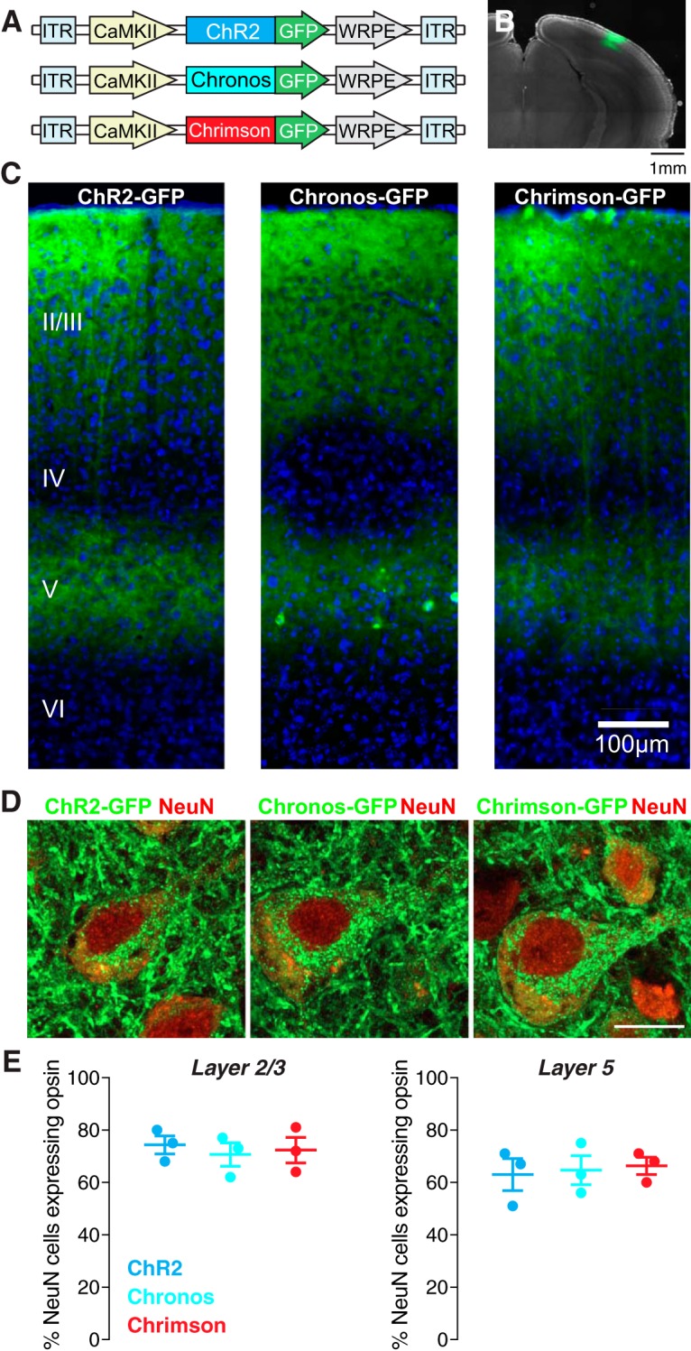
Figure 1.

Expression of three ChR variants in excitatory neurons of the mouse visual cortex. A, AAVs carrying three opsins were injected into primary visual cortex of wild-type mice; ITR, inverted terminal repeat; WRPE, woodchunk hepatitis B virus post-transcriptional element. B, Spread of viral infection in the cortex. Example image showing GFP expression (green) around the area of a cortical injection of AAV5 carrying the Chronos construct. Magnification: 4×. C, ChR2-GFP, Chronos-GFP, and Chrimson-GFP were robustly expressed in excitatory neurons in cortical layers 2, 3, and 5, as confirmed by DAPI staining (blue). Magnification: 10×. D, Example confocal images showing expression of the three ChR variants (green) in layer 5 pyramidal neurons stained for the neuronal marker NeuN (red). Scale bar: 10 μm. Magnification: 64×. E, Quantification of expression in layers 2/3 (left) and 5 (right) for each opsin.
