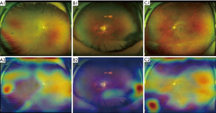
Figure 4.
Ultra-widefield fundus images and corresponding heatmaps showing typical false-positive cases. (A) Retinal pigmented nevus shown on the left of A1 is the reddest region displayed in heatmap A2; (B) Dense retinal hyperpigmentation manifested on the bottom right of B1 is the reddest region visualized in heatmap B2; (C) proliferative vitreous membrane presented on the bottom left of C1 is the reddest region shown in heatmap C2.

