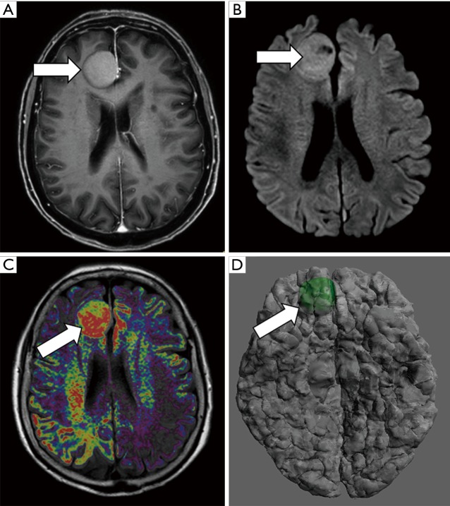
Figure 2.
Meningioma presurgical evaluation. An 81-year-old female with right frontal lobe meningioma. (A) Axial gadolinium-enhanced T1w image shows a large right parasagittal frontal extra-axial lesion consistent with meningioma (arrow). (B) Axial b 1,000 s/mm2 DWI identifies severe restriction of water diffusion within the meningioma which suggests hypercellular lesion (arrow). (C) The fusion of functional information from DWI and morphological information form T1W images demonstrates proper correlation identifying areas of higher hypercellularity within the posterior aspect of the meningioma (arrow). (D) 3D model merging anatomical information from morphological MRI and functional information from DWI allows the evaluation of those areas with a higher restriction of water diffusion within meningioma inside the whole brain anatomy (arrow).

