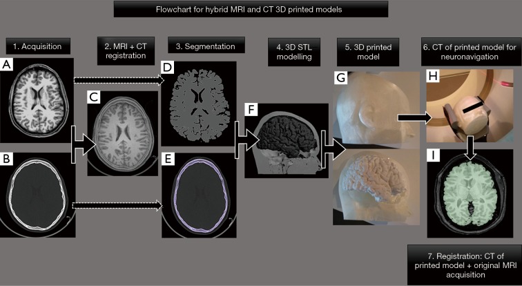
Figure 3.
Flowchart for hybrid MRI and CT 3D printed model for epilepsy pre-surgical planning. A 25-year-old male with a drug-resistant epilepsy candidate for focal thermocoagulative therapy. Neurosurgeons ask for the possibility of obtaining a 3D printed model of the whole patient head including soft tissues, skull, and brain for proper planning of electrodes and guides positioning. Information from both (A) axial 3D-T1W and (B) CT images were used for (C) registration and segmentation of (D) gray matter and (E) bone. (F) Hybrid 3D model in STL format was generated and sent to 3D print obtaining a (G) 3D printed model which included information of both the brain, soft tissues, and skull. (H) The printed brain model has undergone a CT scan which perfectly fitted with its source image form MRI (I). 3D printed model enables neurosurgeons to plan electrodes positioning and surgical entry points before surgery.

