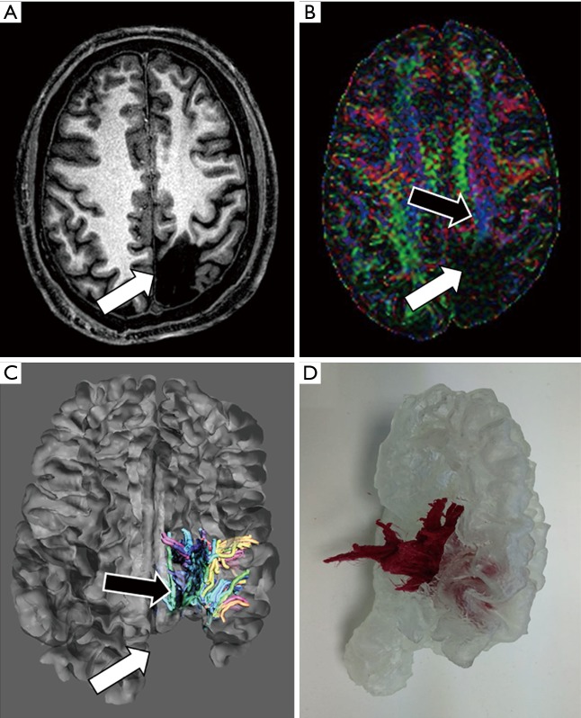
Figure 4.
Presurgical planning of focal epilepsy. A 37-year-old male, with a history of previous surgical left parieto-occipital resection 20 years ago, refers to occipital lobe seizures. Neurologists asked for a presurgical study to minimize the damage of both optical and corticospinal pathways. (A) Axial T1-weighted image shows a large left parieto-occipital area of encephalomalacia (arrow). (B) Color-coded Fraction Anisotropy map, generated from DTI acquisition identifies the involvement of posterior tracts of left corona radiata (white arrow) with apparent preservation of corticospinal tract (black arrow). (C) The fusion of morphological MRI and DTI images allows evaluating the relationships between white matter tracts (black arrow) and post-surgical cavity (white arrow). (D) 3D printed model enables neurosurgeons to address physically the location of white matter tracts for a more secure surgical planning.

