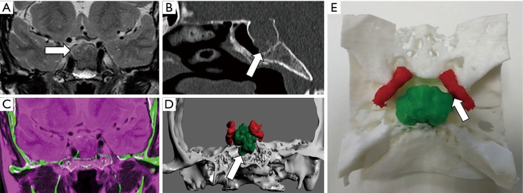
Figure 5.
Presurgical planning of hypophyseal macroadenoma. A 54-year-old female that undergone MRI for headache and vision loss. (A) Coronal T2 TSE shows a large sellar mass that seems to invade sphenoidal sinus (arrow). (B) Sagittal reconstruction of CT confirms the presence of an enlarged sella turcica with disruption of its floor (arrow). (C) Registration of information of skull base structures from CT (green) and data from MRI (magenta) enables an adequate tissue contrast for adenoma and even internal carotids segmentation. (D) Coronal projection of the 3D combined model allows neurosurgeons to properly evaluate the relationship of the hypophyseal mass (codded in green) with sphenoidal sinus, cavernous sinus and even both internal carotids (codded in red), demonstrating disruption of sella turcica floor (arrow). (E) Zenithal view of 3D printed model shows proper correlation with radiological images and 3D virtual model demonstrating contact between macroadenoma and left internal carotid (white arrow).

