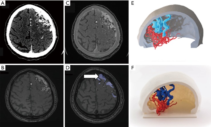
Figure 6.
Brain arteriovenous malformation assessment. A 35-year-old male with left frontal AVM that undergone both CT-angiography and MRI-angiography studies. Information obtained from (A) CT was used for the skull and both arteries and veins segmentation (B). (C) MR-angiography using time of flight (TOF) technique with arterial velocity codification allows accurately segment high flow veins (arrows at Figure 6D) that compose the AVM as well as differentiate them from the brain parenchyma and skull. Combined 3D modeling (E) and 3D printed models (F) provide neurosurgeons and interventional radiologists a comprehensive assessment of AVM and its relationship with the skull.

