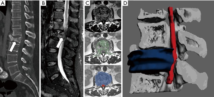
Figure 7.
Spine and spinal cord evaluation. A 42-year-old female with a history of breast cancer is evaluated in an emergency room for lumbar pain and lower limb paresis. (A) Sagittal reconstruction of CT shows L2 vertebral body collapse which seems to invade the spinal canal (arrow). (B) Sagittal STIR sequence confirms the existence of a metastatic fracture with soft tissue component that invades spinal canal conditioning severe stenosis. (C) Registration of information form axial T2-TSE with CT allows to segment vertebral bone (codded in green) and also soft tissue mass (codded in blue) and spinal cord (codded in red). (D) The 3D model enables neurosurgeons to visualize in a single view all the information form both CT and MRI and identify how the soft tissue component (codded in blue) invades the spinal canal and displaces the spinal cord.

