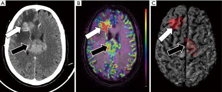
Figure 8.
Multimodality assessment of a CNS tumor. A 73-year-old female with changes in behavior and cognitive impairment undergone (A) contrast-enhanced CT that shows two large hyperenancing masses at right frontal lobe (white arrow) and within the corpus callosum (black arrow). (B) MRI was performed using ASL technique for assessment of tumor blood flow before guide biopsy to avoid the administration of gadolinium. The fusion of morphological MRI sequence (FLAIR) and functional MRI sequence (ASL) allows neuroradiologists to characterize the right frontal lesion as a potentially higher grade compared with the corpus callosum lesion as the former shows higher blood flow values (white arrow) compared with the latter (black arrow). (C) The register of morphological MRI and functional (ASL) MRI data allows the generation of a 3D model in a single file with all the information needed for a proper planification of brain biopsy.

