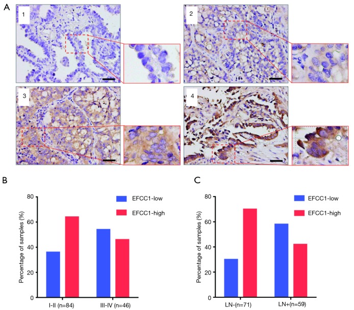
Figure 4.
EFCC1 expression in clinical lung ADC samples with TMA. (A) Representative images of different immunohistochemical staining intensities of EFCC1 in clinical lung ADC tissues. 1, negative staining; 2, weak staining; 3, moderate staining; 4, strong staining. Scale bar =20 µm. (B) The proportion of low EFCC1 expression was significantly higher in lung ADC patients at stage III–IV (54.3%) than those at stage I–II (35.7%) (P=0.040). (C) The proportion of low EFCC1 expression was significantly higher in lung ADC patients with lymph node metastasis (57.6%) than those without lymph node metastasis (29.6%) (P=0.001). EFCC1, EF-hand, and coiled-coil domain-containing; ADC, adenocarcinoma; TMA, tissue microarray; TNM, tumor-node-metastasis; LN, lymph node.

