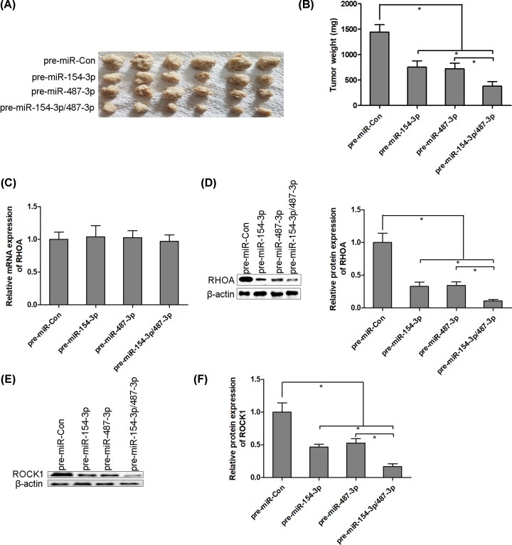
Figure 6. miR-154-3p and miR-487-3p repress thyroid cancer K-1 cell growth in vivo.
Thyroid cancer K-1 cells (1 × 10⁷ cells per 0.1 ml) were established to steadily express miR-154-3p, miR-487-3p or miR-154-3p/487-3p. K-1 cells were implanted subcutaneously into 4-week-old BALB/c nude mice, and tumor growth was evaluated at week 4 after K-1 cells implantation (A and B). The mRNA and protein levels of RHOA are detected using RT-PCR and Western blotting, respectively, in solid tumors of K-1 cells transplanted nude mice (C and D). The protein levels of ROCK1 are detected using Western blotting in solid tumors of K-1 cells transplanted nude mice (E and F); * P < 0.05.

