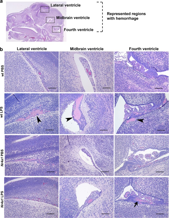
Fig. 4. Comparison of cerebral pathology observed in wt and Nr4a1 KO embryos.
a H&E stained cross-section of an E15.5 brain, highlighting the regions (boxed areas) where injury in the form of hemorrhage was mainly observed. b Representative micrographs of hemorrhage in the respective areas described in a for group 1 (PBS vs. LPS). Hemorrhage was predominant in wt brains exposed to LPS (arrowheads) and to a lesser extent in Nr4a1 KO brains also exposed to LPS (arrow). Scale bars denote 100 µm.

