
Figure 4.
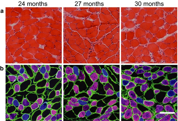
Representative images for histological section of the diaphragm muscle (DIAm) for 24, 27, and 30 month old C57BL/6 × 129 mice: hematoxylin and eosin stain (a); immunohistochemistry based on DIAm myosin heavy chain (MyHC) isoform composition (b): blue—type I fibers, purple—type IIa fibers, and black type IIx and/or IIb fibers. Note lack of evident fiber damage and differences in fiber dimensions across fiber types without obvious changes into very old age. Laminin immunoreactivity is shown in green. Scale bars are 50 µm