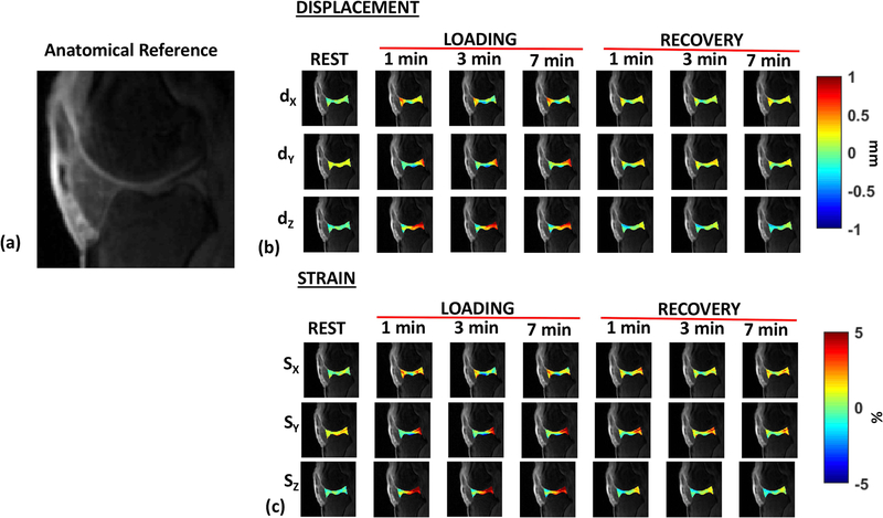
Figure 4.
Representative dynamic displacement and strain maps. (a) shows the anatomical reference for the cartilage joint (b) shows the displacement (dX, dY, and dZ) for the rest, loading condition (1, 3 and 7 minutes into the loading phase), and recovery condition (1, 3 and 7 minutes into the recovery phase). (c) shows the corresponding strain (SX, SY, and SZ) along the X, Y and Z directions for the rest, loading and recovery phases.

