
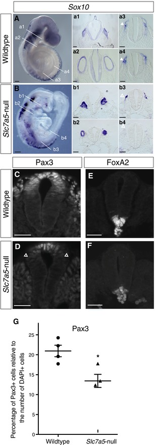
Figure 3. Slc7a5‐null embryos exhibit altered expression of neural crest genes.
-
A, BSox10 in situ hybridisation in (A) wild‐type and (B) Slc7a5‐null E9.5 embryos (n = 4 each) with TSs of (a1, b1) the hindbrain at level of trigeminal ganglion V, (a2, b2) otic vesicles and the neural tube at more posterior levels (a3, a4, b3, b4). Scale bars 200 µm, except sections a1–b4, 100 µm. Closed white arrowheads in (a3, a4) indicate neural crest, and open white arrowheads indicate depleted neural crest in (b3) and position where neural crest should be in (b4).
-
C–FImmunofluorescence on TS caudal spinal cord of E9.5 wild‐type littermates and Slc7a5‐null embryos; Pax3 and FoxA2 were used as indicators for dorso‐ventral organisation in (C, E) wild type, n = 2 (for FoxA2) and n = 4 (For Pax3) and (D, F) Slc7a5‐null, n = 3 (for FoxA2) and n = 3 for Pax3) neural tube. Arrowheads indicate border of Pax3 expression domain. Scale bars 50 µm.
-
GPercentage of Pax3‐expressing cells was determined by counting these cells and all DAPI‐labelled nuclei in the neural tube and comparison made between wild type (four embryos, six sections each) and Slc7a5‐null (three embryos, six sections each), each dot represents the average for one embryo, unpaired t‐test, *P = 0.018 (see original source data). Error bars indicate SEM.
Source data are available online for this figure.