Figure 3.
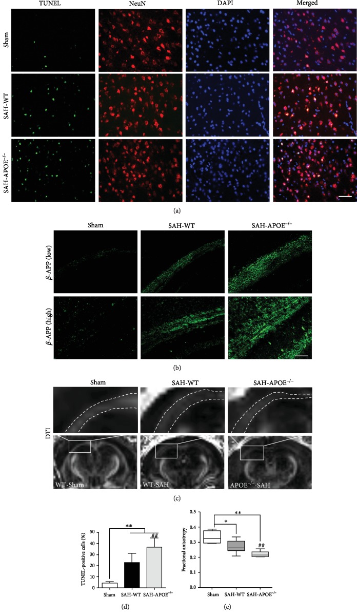
APOE deficiency aggravated neuronal damage in the early phase of SAH. (a) Costaining of TUNEL and NeuN showing apoptotic neurons. (d) SAH induced neuronal apoptosis in the cortex (∗∗P < 0.01, compared to sham, n = 5), while the number of apoptotic neurons of APOE−/− mice was more than that of WT mice (##P < 0.01). (b) APOE deficiency aggravated β-APP accumulation in the white matter after SAH (n = 5). Low magnification (200x), high magnification (400x). (c) DTI showing FA decrease after SAH. (e) SAH induced FA decrease (∗P < 0.05, ∗∗P < 0.01, compared to sham, n = 4), while the FA of APOE−/− mice in the white matter was lower than that of WT mice (##P < 0.01). Bar = 50 μM.

