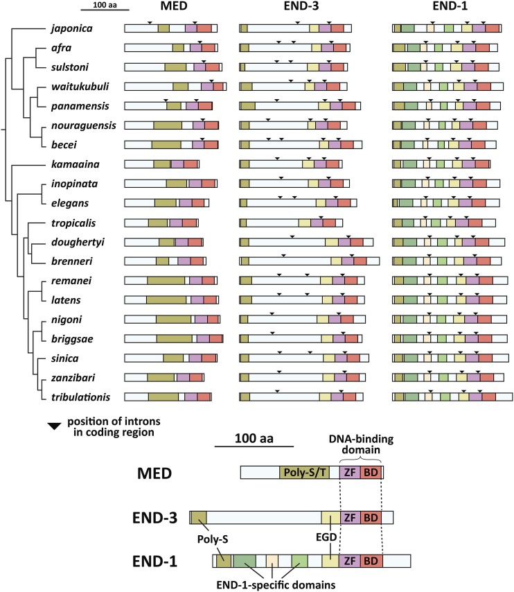
Figure 8.
Conserved MED and END protein domains. The top part of the figure shows the MED, END-3 and END-1 protein structures with conserved domains in colored regions. Triangles represent the positions of introns in the coding regions as shown in the gene models in Fig. 4A. The bottom of the figure shows the names of the domains, which are shown at the amino acid level in Figs. 9 and 10. The MED orthologs have a variable region high in serine and threonine (Poly-S/T), while END-1 and END-3 share an amino-terminal polyserine domain (Poly-S) of variable length and an Endodermal GATA Domain (EGD). The END-1 orthologs share three additional regions not found in END-3. The species are arranged after the phylogeny in (Stevens et al. 2019).

