
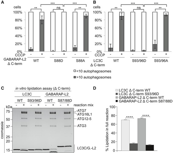
Figure 6. Phospho‐mimetic Δ C‐terminal LC3C or GABARAP‐L2 cannot localize to autophagosomes because they cannot be lipidated.
-
A, BU2OS cells were transfected with GFP‐GABARAP‐L2 Δ C‐terminal (A) or GFP‐LC3C Δ C‐terminal (B) WT or mutants and HA‐Parkin. Mitophagy was induced by the addition of 40 μM CCCP for 3 h. GFP‐expressing cells were counted and segregated into classes with greater and less than 10 autophagosomes per cell. Data are presented as mean ± SD, n = 3 biological replicates, **P < 0.01, ***P < 0.001 as analyzed by Student's t‐test.
-
CIn vitro lipidation reactions containing 10 μM LC3C WT, LC3C S93/96D, GABARAP‐L2 WT, or GABARAP‐L2 S87/88D incubated with or without reaction mix (0.5 μM hATG7, 1 μM hATG3, 0.25 μM hATG12‐ATG5‐ATG16L1, 3 mM lipid (sonicated liposomes composed of 10 mol% bl‐PI, 55 mol% DOPE, and 35 mol% POPC), 1 mM DTT, and 1 mM ATP) were incubated at 30°C for 90 min. The reactions were analyzed by SDS–PAGE and visualized by Coomassie blue stain.
-
DThe extent of lipidation in (C) was quantified and plotted as percentage of total protein (conjugated and unconjugated). Data are presented as mean ± SEM, n = 3 biological replicates, ****P < 0.0001, as analyzed by one‐way ANOVA followed by Bonferroni's multiple comparison test.
Source data are available online for this figure.