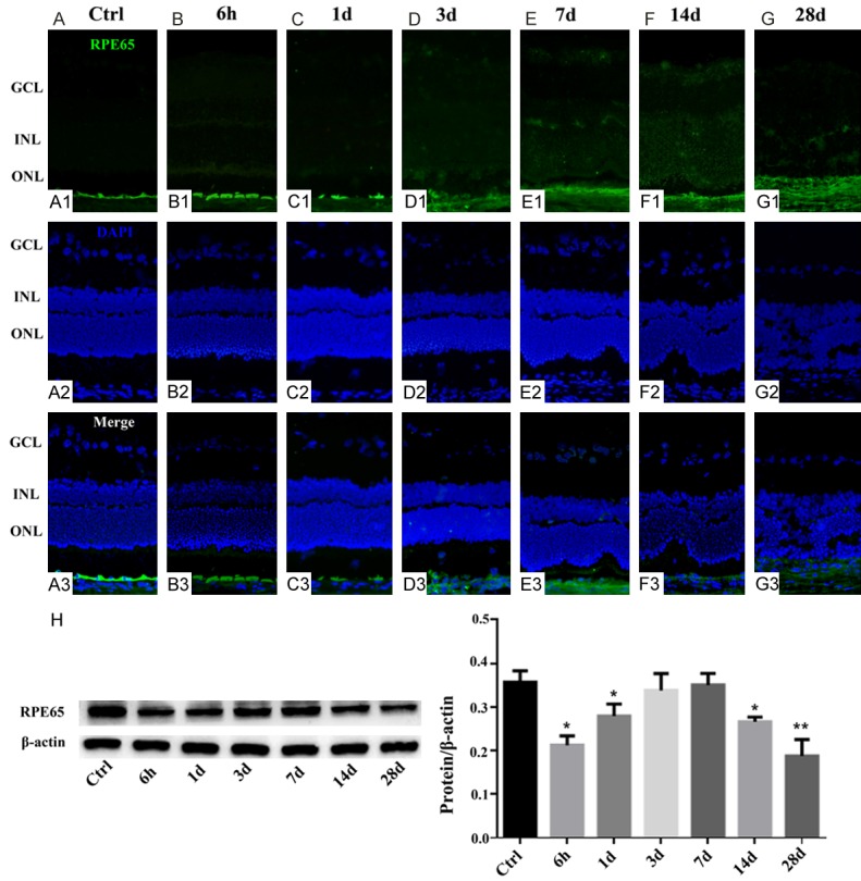
Figure 2.

Effect of NaIO3 on RPE65 expression in the RPE cells in retinas. Immunocytochemical staining for RPE65 in the retinas of NaIO3-treated and control rats. Control healthy retina with no signs of injury (A). The RPE cell was degenerated at 6 h post NaIO3 injection (B), and this was particularly obvious at 1 day PI (C). Histological assessment of the retinas at 3-7 days PI revealed regeneration of the RPE structure at the sites of injury (D, E). Then the remaining RPE structure could not be detected indicating direct signs of local injury to the RPE (F, G). Western blotting was used to confirm RPE65 expression in the groups (H). The graph shows the ratio of RPE65 to β-actin in each group. RPE65 expression was reduced in other NaIO3 group but increased significantly 3 and 7 days PI (*, P < 0.05; **, P < 0.005).
