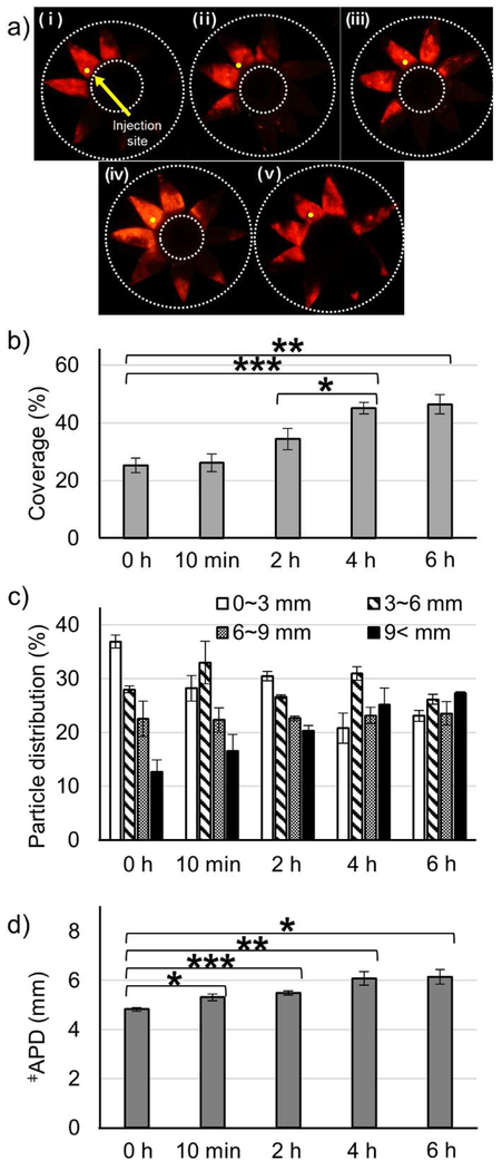
Figure 3.
Effect of collagenase incubation time on microparticle delivery in the SCS. Collagenase formulation (0.5 mg/mL collagenase with 3 mM CaCl2) was injected into the SCS of rabbit eyes ex vivo and incubated at 37 °C for different lengths of time. Representative fluorescence micrographs (a), particle delivery coverage (b), particle distribution (c), and APD (d) of red-fluorescent microparticles after injection into the SCS followed by different incubation times: (i) 0 h (ii) 10 min (iii) 2 h (iv) 4 h (v) 6 h. ǂAverage particle distance. Graphs show mean ± SEM (n=3 in (i, ii, iii, and v) and n=6 in (iv)). *,**,*** indicate significant difference (one tailed t-test, p<0.05,0.01,0.005, respectively).

