Figure 5.
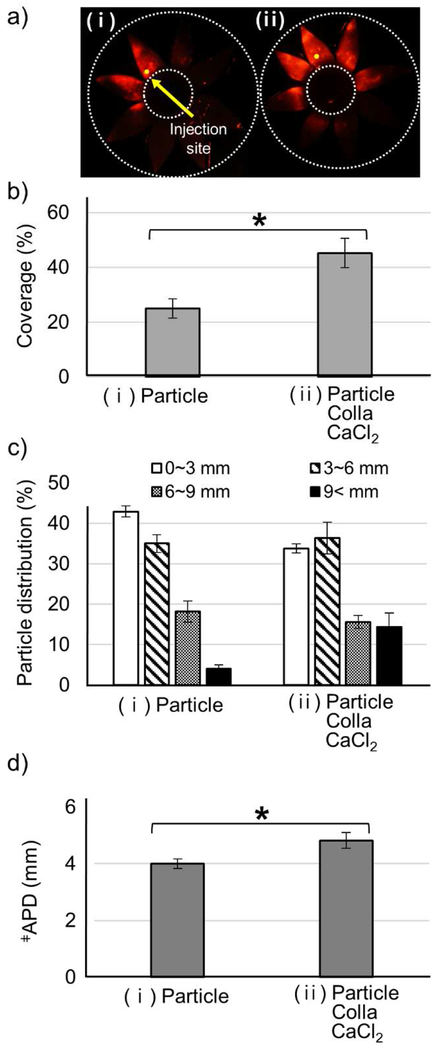
In vivo microparticle delivery with collagenase. Collagenase formulation (0.5 mg/mL collagenase with 3 mM CaCl2) was injected into the SCS of rabbit eyes in vivo. Representative fluorescence micrographs (a), particle delivery coverage (b), particle distribution (c), and APD (d) of red-fluorescent microparticles in the SCS determined 4 h after the injection of (i) control solution containing microparticles in HBSS (Particle) and (ii) collagenase formulation with microparticles (Particle Colla CaCl2). ǂAverage particle distance. Graphs show mean ± SEM (n=3). * indicates significant difference (one tailed t-test, p<0.05).

