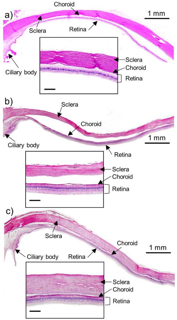
Figure 6.
Representative histological images of rabbit eye tissue collected 4 h after SCS injection in vivo. (a) Untreated eye, (b) eye injected with control solution containing microparticles in HBSS and (c) eye injected with collagenase containing microparticles in HBSS. Tissue was chemically fixed and stained with hematoxylin-eosin (H&E). Scale bars of the insets are 200 μm.

