Figure 1.

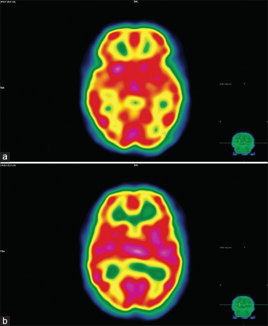
(a and b) Hexamethylpropyleneamine oxime single-photon emission computed tomography shows moderate hypoperfusion of the primary visual cortex, expanding to the temporo-occipital junction bilaterally but predominantly on the right side

(a and b) Hexamethylpropyleneamine oxime single-photon emission computed tomography shows moderate hypoperfusion of the primary visual cortex, expanding to the temporo-occipital junction bilaterally but predominantly on the right side