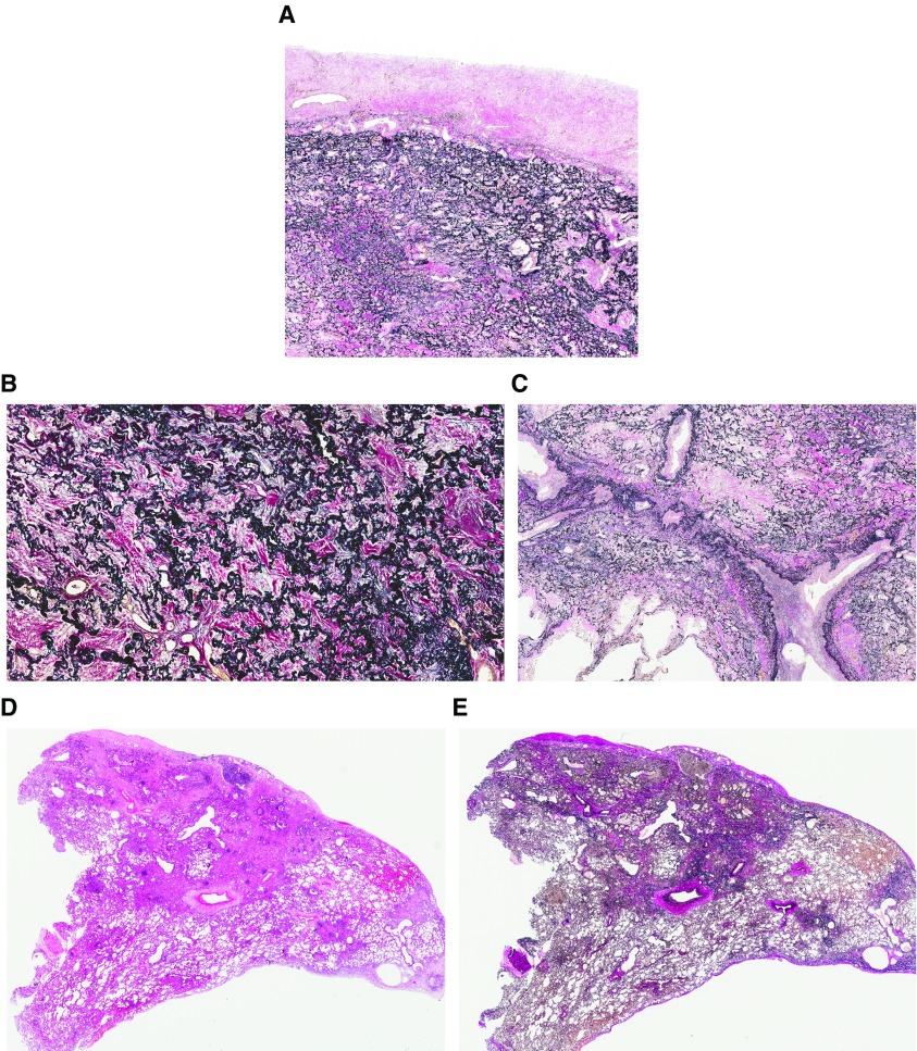
Figure 4.
(A) Elastic van Gieson staining showing a combination of visceral pleural fibrosis and intraalveolar fibrosis with elastosis (IAFE). IAFE comprising dense collagenous fibrosis fills the alveolar spaces, whereas the residual alveolar walls are highlighted by elastin deposition. (B) At higher power, the alveolar parenchyma can appear “petrified” by the fibrosis, its architecture highlighted by abundant elastosis. (C) Intimal fibrosis within the pulmonary vasculature, particularly the pulmonary veins, is a common finding in pleuroparenchymal fibroelastosis. (D and E) Although IAFE predominates in the subpleural lung parenchyma, it may extend into the deeper lung, typically around interlobular septa and bronchovascular bundles, as shown to the right of both panels. Elastic van Gieson staining is shown in A, B, C, and E; hematoxylin and eosin staining is shown in D.

