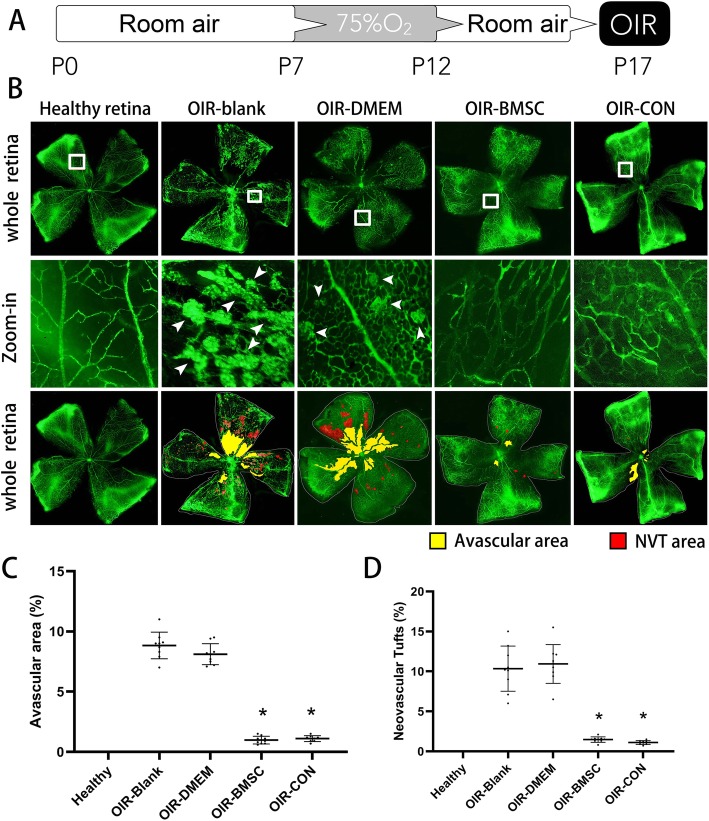
Fig. 1.
Neovascularization was inhibited after BMSC or Conbercept injection. a Schematic process of establishing an OIR animal model by exposure to 75% oxygen from postnatal day 7 (P7) to P12 and return to room air with or without intravitreal injection. b P17 retinal flat-mount revealed neovascular tufts (NVT, arrowheads) and avascular areas developed after high oxygen exposure (OIR-blank and OIR-DMEN) but shrinked following BMSC (OIR-BMSC) or Conbercept (OIR-CON) injection. c Avascular areas were presented as percentages to the whole retina; both BMSC and Conbercept injection significantly reduced avascular areas. d Similar quantification was calculated in regard to the neovascular tufts (NVT) areas. *, P < 0.05 versus OIR-blank; n = 9

