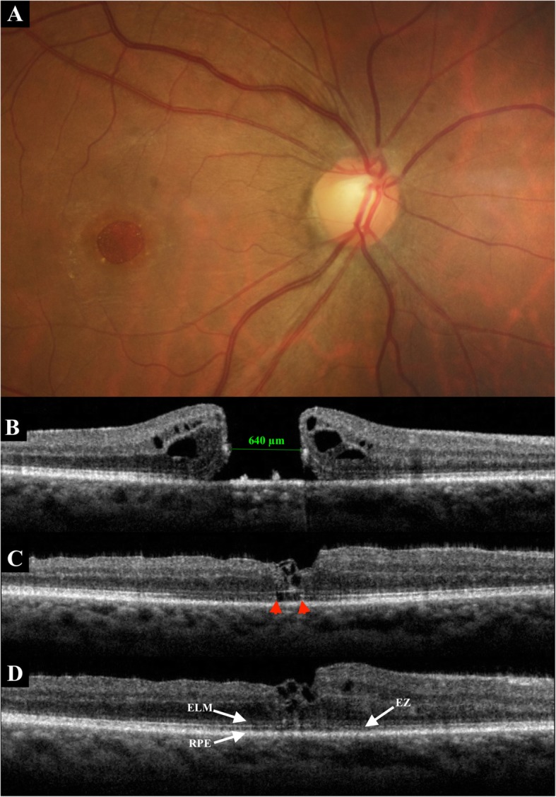
Fig. 1.

A 61-year-old man presented with idiopathic large MH. a. Fundus photography before surgery. b. Baseline SD-OCT. The minimum diameter was 640 μm. Preoperative BCVA was 0.04. c. SD-OCT at 1 month postoperatively. Inverted ILM flap technique was performed, with a W-shape closure (irregular closure) of the MH. SD-OCT showed the foveal hyperreflective tissue corresponding to a coiling of the flap. Disruption of the EZ (between red arrowheads) remained. The ELM line was almost complete. BCVA was 0.16. d. Six months after surgery, SD-OCT showed a persistent W-shape closure of the MH with a wrapped flap. There was a complete restoration of the ELM and EZ lines. The BCVA increased to 0.5
