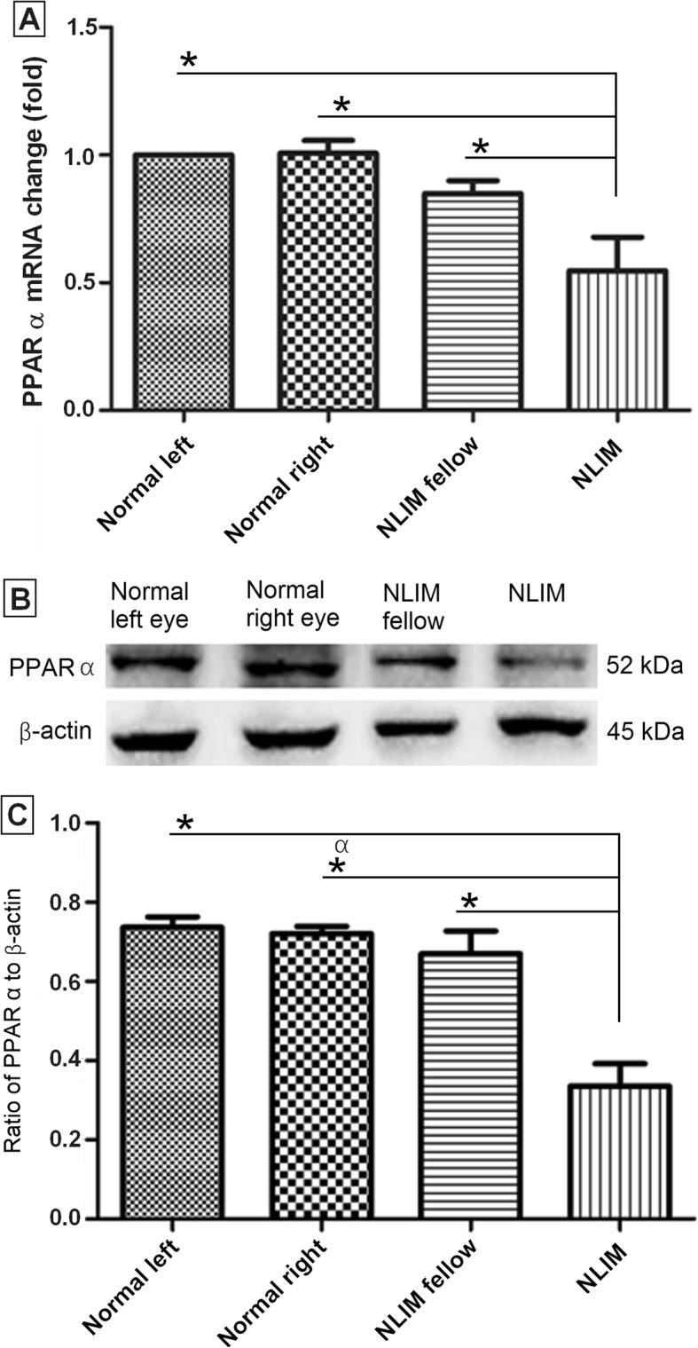
Fig. 6.

Determination of PPAR α at mRNA and protein levels in normal control and NLIM guinea pig sclera. Using pooled samples, quantitative PCR (a) and western blotting (b) analyses were done, and histogram analysis was carried out for western blotting (c). Data were presented as mean ± SD for mRNA and protein expressions (n = 6 for each group) and *P < 0.05
