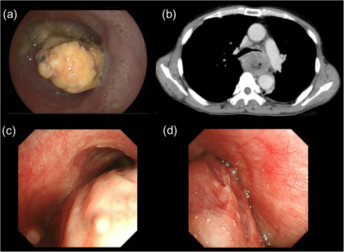
Fig. 1.
a Esophagogastroduodenoscopy revealed that the esophageal lumen was filled with necrotic components from the primary esophageal cancer; b computed tomography (CT) revealed that the left bronchus was compressed by the primary esophageal tumor components; c bronchoscopy revealed that the tracheal lumen at the carina level was deformed by the primary esophageal tumor components; d bronchoscopy revealed that the left bronchus presented with an almost complete obstruction due to compression by the primary esophageal tumor components

