Fig. 2.

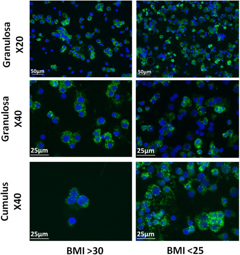
Fluorescent microscopy of granulosa and cumulus cells Nile Red staining. Fluorescent microscopy of granulosa and cumulus cells. Lipid droplets (green) are stained with Nile Red and cell nuclei (blue) are stained with DAPI

Fluorescent microscopy of granulosa and cumulus cells Nile Red staining. Fluorescent microscopy of granulosa and cumulus cells. Lipid droplets (green) are stained with Nile Red and cell nuclei (blue) are stained with DAPI