Figure 6.
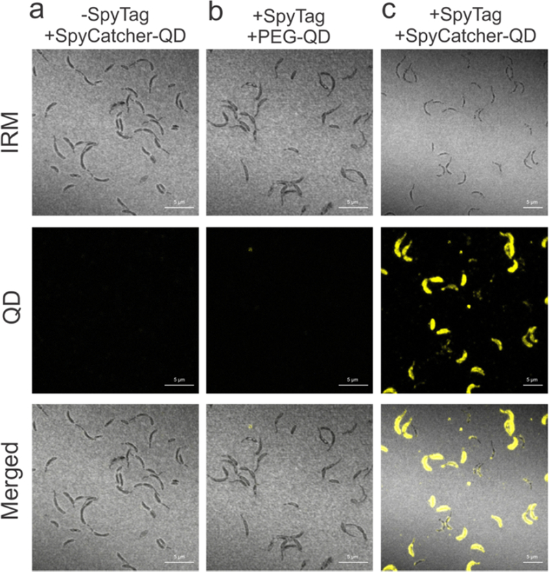
Engineered RsaA assembles inorganic nanocrystals on the C. crescentus cell surface. (a–c) Interference reflection microscopy (IRM) and confocal fluorescence images of C. crescentus cells incubated with QDs. Cells expressing (a) wild-type RsaA incubated with SpyCatcher-QDs and (b) expressing RsaA690:SpyTag incubated with PEG-QDs. (c) Cells expressing RsaA690:SpyTag incubated with SpyCatcher-QDs show QD fluorescence along the cell surface, indicating specific assembly of SpyCatcher-QDs by the engineered strain. Scale bar = 5 μm.

