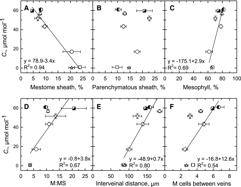
Figure 4.
The relationship between leaf structural parameters and C* in eight Neurachninae species. Values determined from light microscopic images of planar cross sections. Percent tissue values were calculated as the area of a given tissue in a leaf image divided by the sum of the areas of MS, PS, and M tissues in the same image, times 100% (Supplemental Fig. S2). A, Percent of image area composed of MS tissue. B, Percent of image area composed of PS tissue. C, Percent of image area comprised of M tissue. D, The ratio of M tissue area to MS tissue area in planar cross section. E, Mean distance between veins. F, The number of M cells between veins. Mean ± se; n = 3 plants per species; 10 images per plant. See Supplemental Table S1 for mean values and statistical tests. Significant linear regressions at P ≤ 0.05 are shown. N. alopecuroidea (◑), N. annularis (▽), N. lanigera (◇), N. minor (○), N. munroi (⬜), N. muelleri (☆), N. queenslandica (△), and T. mitchelliana (◪).

