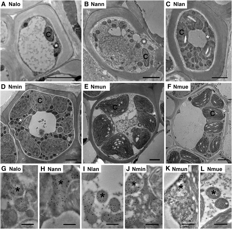
Figure 5.
TEM images of MS cells of six Neurachne species. A to F, Low magnification images illustrating ultrastructure. G to L, Immunogold labeling of GLDP in MS mitochondria. Immunogold labeling of GLDP is indicated by black dots in mitochondria. Nalo, N. alopecuroidea; Nann, N. annularis; Nlan, N. lanigera; Nmin, N. minor; Nmun, N. munroi; Nmue,N. muelleri; C, chloroplast; *, mitochondrion. Scale bars = 2 μm (A–F); 500 nm (G–L).

