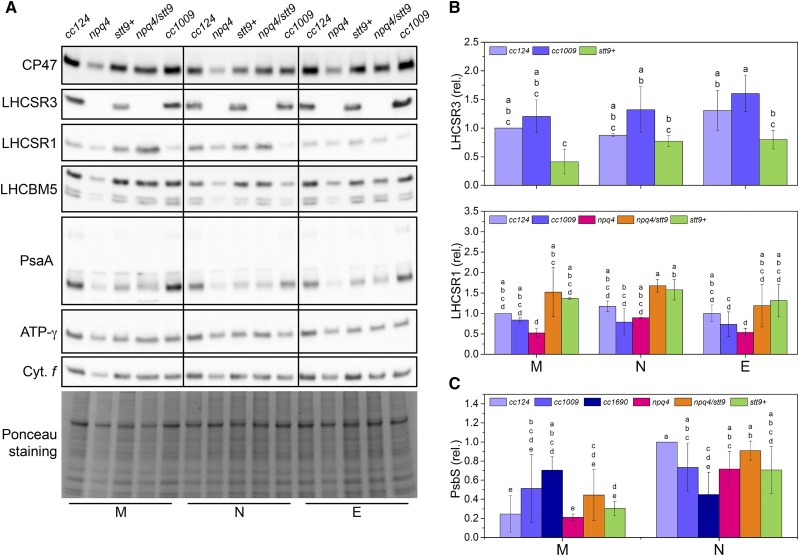
Figure 3.
The main proteins involved in photoprotection are present in Chlamydomonas cells throughout the day. M, N, and E denote samples isolated at the onset of illumination (morning), at the peak light intensity (noon), or at the end of the light period (evening), respectively. Shared lowercase letters between means denote the absence of a statistically significant difference (P < 0.05; see “Materials and Methods” for details). A, Immunoblot analysis with antibodies against the subunits of major photosynthetic complexes assessed throughout the day-night cycle. Whole-cell extracts were loaded on an equal-protein-content basis. Ponceau staining of the membrane as a loading control is shown at the bottom. B, Quantification of the LHCSR content throughout the day-night cycle. The means ± sd were calculated for three biological replicates. C, Quantification of the PsbS content throughout the day-night cycle. The means ± sd were calculated for three biological replicates.

