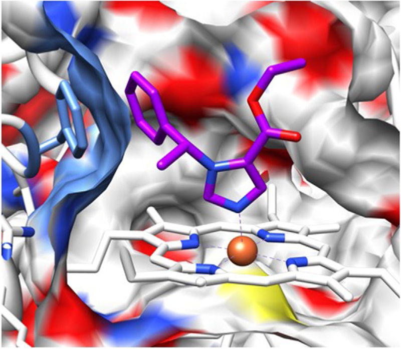
Fig. 2.
Homology model of 11β-hydroxlase with etomidate computationally docked. A cross section through the surface of the active site cavity is shown. Etomidate is depicted in stick representation with blue nitrogens, red oxygens, and purple carbons. The heme iron is orange and coordinates with etomidate’s aromatic imidazole nitrogen. The authors thank Drs. Keith W. Miller and Sivananthaperumal Shanmugasundararaj for producing this figure.

