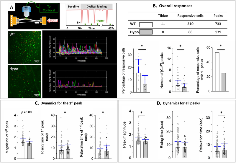
Figure 1.
Osteocytes of Hypo murine tibiae showed impaired intracellular calcium [Ca2+]i response to mechanical loading. (A) Freshly dissected murine tibiae were subjected to axial cyclic loading (8 N peak load), and intracellular calcium [Ca2+]i of osteocytes beneath the anterior-medial surface were imaged using confocal microscopy and trigger signals inserted between loading cycles. Representative images of osteocytes (green, left panel) and normalized [Ca2+]i traces of individual cells (right panel) are shown for tests using WT and Hypo tibiae. (B) The overall responses included the percentage of responsive cells over the total number of cells, the number of [Ca2+]i peaks (averaged for all responsive cells), and the percentage of responsive cells with multiple [Ca2+]i peaks. (C) Dynamics for the first peak included the magnitude, rising time, and relaxation time. (D) Dynamics for all peaks included the magnitude, rising time, and relaxation time. Mean, standard deviation, and individual data points are shown. * indicates statistical significance p < 0.05 between WT and Hypo using Mann–Whitney U or Chi-square tests.

