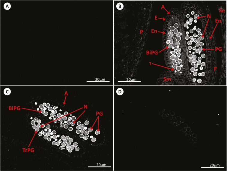
Fig. 3.
Immunolocalization of GSH by antibody-associated fluorescence microscopy in Arabidopsis anthers at stage 14. (A) Negative control; (B) WT; (C) roGFP2; (D) cad2-1/roGFP2. A, anther; BiPG, bicellular pollen grain; E, epidermis; En, endothecium; N, nucleus; P, petal; PG, pollen grain; Se, sepal; Sm, stamen; T, tapetal layer; TrPG, tricellular pollen grain.

