Figure 4.

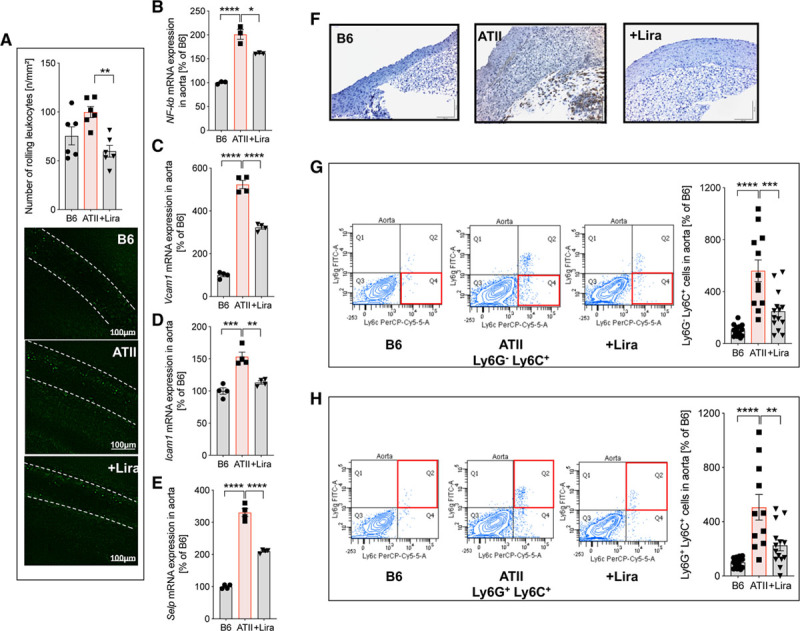
Liraglutide (Lira) ameliorates ATII (angiotensin II)-induced inflammation of the vasculature by prevention of Ly6G−Ly6C+- and Ly6G+ Ly6C+ cell infiltration to the vascular wall. A, In wild-type mice (C57BL/6J), rolling of leukocytes at the vascular wall (V. femoralis) was quantified by intravital microscopy. Nucleated cells were visualized by staining with acridine orange. Representative images at day 7 are shown below the quantification bar graph. Representative videos of intravital microscopy experiments are available in the online-only Data Supplement. B–E, mRNA expression of genes encoding for nuclear factor-κb (NF-κb) and adhesion molecules vascular cell adhesion molecule-1 (Vcam1), intercellular adhesion molecule-1 (Icam1), and P-selectin (Selp) in aorta were measured by quantitative reverse transcription polymerase chain reaction. F–H, Vascular inflammation was further characterized by immunohistochemistry and flow cytometry of aortic tissue. Representative pictures of 3–4 independent aortic Ly6 (Ly6B.2) immunohistochemical stainings (F; for IgG isotype control see Figure V in the online-only Data Supplement) and flow cytometry of Ly6G−Ly6C+ inflammatory monocytes and Ly6G+Ly6C+ neutrophils in aortic cell suspensions (G and H) are shown. Representative original plots of Ly6G−Ly6C+ and Ly6G+Ly6C+ cells are shown beside the quantification bar graph (for gating strategy, see Figure IV in the online-only Data Supplement). One-way ANOVA and Bonferroni multiple comparison test; N=6 (A), 3 (B–E) and 12-14 (G and H) animals per group. Data are means±SEM. *P<0.05; **P<0.01; ***P<0.001; ****P<0.0001.
