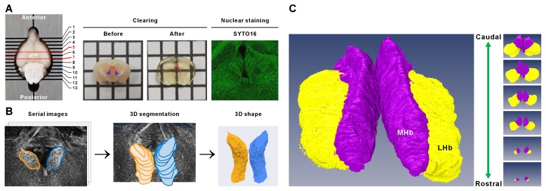
Fig. 1.
Data acquisition and 3D reconstruction of mouse habenula. (A) Brain was removed from the head and cut into a 2-mm thick coronal section (line 5 and 7) including the habenula (Hb) using coronal mouse brain matrix at 1-mm intervals. Dissected brain was processed with 3 hours of ACT (before and after ACT shown) and nuclear staining (SYTO16) was performed. (B) The assessment of volume and shape in the Hb: Brain samples were processed following the steps depicted in serial image acquisition and 3D segmentation to obtain 3D volumetric models of the Hb. (C) Dorsal view of representative volumetric reconstructions of the Hb (purple, MHb; yellow, LHb) with rostral to the top (caudal).

