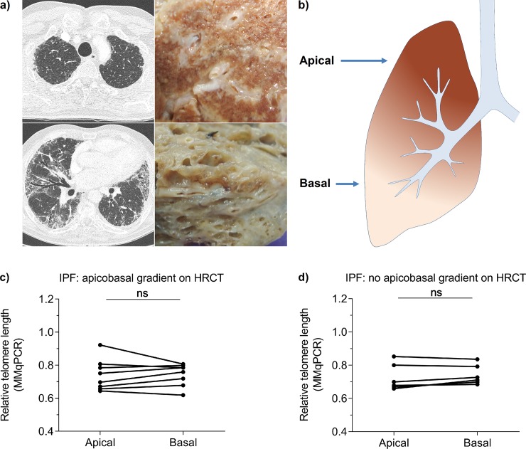
Fig 3. Telomere shortening is not associated with apical or basal localisation in whole explant lung.
(a) HRCT on the left and formalin fixed explant images of IPF lung with an apicobasal fibrotic gradient on the right. The top and bottom figures represent apical and basal locations respectively, corresponding to (b) the schematic picture. (c, d) Apical and basal lung telomere length comparison of (c) 8 lungs with an apicobasal gradient and (d) 7 lungs without apicobasal gradient measured by MMqPCR. Samples belonging to the same person are connected. Wilcoxon matched-pairs signed rank tests showed no differences in telomere length between lung sections.

