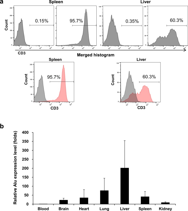
Fig 7. FACS staining and Alu PCR in mouse organs.
a Post sacrificing the mice on day 3 after Jurkat/CAR T-cells injection, graphs of FACS staining for liver and spleen tissues of mice were plotted against the control group. b Alu PCR analysis data of mouse blood, brain, heart, lung, liver, spleen, kidney and gut tissues obtained from sacrifice three days after Jurkat/CAR T-cells injection.

