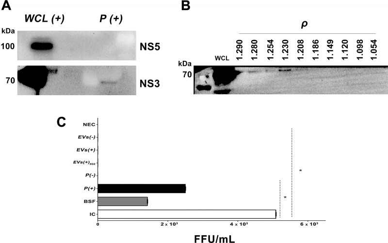
Fig 3. EVs of infected macrophages contain the NS3 protein of DENV-2 but not infectious material.
(A). 10 μg/μL of an extract of infected cells (WCL (+)) or 15 μg/μL of P(+) were processed by SDS-PAGE and WB to detect the DENV NS5 (105 kDa) or NS3 (70 kDa) proteins. These samples were positive only for NS3 protein. WB representative of two tests per condition is shown. (B). The EVs of infected cells with densities (ρ) between 1.23 to 1.28 g / mL were positive for the NS3 protein, corresponding to ABs (H3 positive). As control we used WCL (+). (C). After UC, the EVs of the uninfected and infected macrophages were processed by immunoprecipitation (IP) and the resulted vesicles from uninfected (EV (-)) or infected (EV (+)) pellets were inoculated on LLC-MK2 cells and then foci formation were evaluated by immunoperoxidase at 72 h p.e. As controls the following conditions were evaluated: non-exposed cells (NEC); LLC-MK2 cells infected with DENV-2 MOI: 0.2 (IC); cells inoculated with beads free supernatants (BFS) obtained after the IP process; cells inoculated with the P (-) or P (+); vesicles of U937 infected cells, purified by IP and treated with 4G2 antibody (EVs (+) 4G2) or vesicles of U937 non-infected cells (EVs(-)) purified by IP. Viral antigen was detected when LLC-MK2 cells were exposed to DENV-2, BFS, and P (+). Mean of focus-forming units of two independent experiments by duplicate is shown. Data was analyzed using the Kruskal–Wallis test, followed by a Mann–Whitney test, with p<0.05.

